Antibody hypothesis of where they are and who secretes them. After the IgD and IgM which are non discriminating have failed to work the immune system becomes more specific.
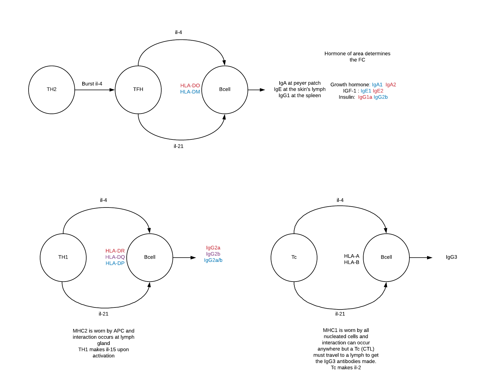
T cells have been educated to know self antigens from the inside of cells. HLA mailboxes hold up suspect foreign pieces to the T cells.
Tcellreceptors match up with HLA mailboxes of Bcells. If the piece held in the mailbox is foreign then the T cell secretes il-4 and il-21 to stimulate proliferation and antibody secretion by the B cells.
The B cell's location and T cell partner determines what antibody is secreted. Note that APC, antigen presenting cells, can kickstart the cytokine process but only B cells secret antibodies.
Th2 cells become Tfh (the follicular helper T cells) who tell B cells to secrete antibodies based on location:
the gut (mucosal area/ peyer patches) IgA
the spleen (blood/body) IgG1
the skin's lymph IgE
Th2 to Tfh
https://www.ncbi.nlm.nih.gov/pubmed/19380637
Th1 cells who match up with B cells or APC with MHC2 (HLA-d family) stimulate B cells to make IgG2.
There are 3 forms of IgG2:
IgG2a which appears with the flu virus
IgG2b which appears with reoviruses
IgG2a/b which would bind proteins etc.
These may match up with the HLA-d
HLA-dr flu virus
HLA-dq reovirus
HLA-dp bacterias
Disulfide bonds are highly charged and would attract nucleotides.
IgG1 has only 2 disulfide bonds which does not attract nucleotides (TFH secreted)
IgG2 has 4 disulfide bonds which would be strong enough to lure nucleotides. (TH1 secreted)
The heterodimer form IgG2a/b is asymmetric and would not bind nucleotides. (bonds don't match up)
Note that there will be overlap with infections because when bacteria are broken open their inner DNA/RNA would be exposed.
There are 2 forms of IgA (TFH secreted)
IgA2 triggered with TGF-b1 is IgA dimer of 2 held together by a protein with tons of disulfide bonds. Which would attract nucleotides. Imagine a huge clump formation of antibodies and viruses which is easy for the immune system to dispose of.
IgA1 triggered by il-5 is not a dimer and does not attract nucleotides.
Which leaves the Tc and B cell with MHC1 (HLA-A/B) which bind the viruses of the mitochondria or the nucleus. These B cells would produce IgG3. This antibody has 11 disulfide bonds which would be strongly attractive for nucleotides.
the CTL in the secondary lymph (having seen the antigen HLA at the site of infection)
https://www.ncbi.nlm.nih.gov/pmc/articles/PMC5788973/
https://www.ncbi.nlm.nih.gov/pubmed/12524535
https://www.ncbi.nlm.nih.gov/pubmed/17513747/
https://www.ncbi.nlm.nih.gov/pubmed/11777979
How do the TFH's B cells know where they are and which antibody to make? Since there are 3 different antibodies at 3 different locations they must sense where they are.
Hypothesis: Insulin, growth hormone, and insulin-like growth factor coordinate which antibody is produced.
1. The mucosal membrane/ peyer patches
Growth hormone deficiency is connected to IgA deficiency
https://www.ncbi.nlm.nih.gov/pubmed/7527197
Growth hormone and the intestine
https://www.ncbi.nlm.nih.gov/pubmed/10905374
2. The skin interface
Insulin like growth factor IGF-1 and IgE
https://www.ncbi.nlm.nih.gov/pubmed/8046348
IGF-1 and skin
https://www.ncbi.nlm.nih.gov/pubmed/16537911
3. The spleen
insulin and IgG2
https://www.ncbi.nlm.nih.gov/pubmed/2659538
Note that the tail of the pancreas is at the spleen and the blood supply is shared
HLA hypothesis
http://angelabiggs.blogspot.com/2016/10/hla-location-hypothesis.html
The germinal Bcells use HLA-DO and HLA-DM.
https://www.ncbi.nlm.nih.gov/pmc/articles/PMC2193692/
This would explain why there are 2 forms of Antibody from the germinal center. The HLA-DO is similar to HLA-DR which binds viruses.
IgE1 and IgE2
https://www.ncbi.nlm.nih.gov/pubmed/9257792
IgG1a and IgG1b
http://www.pnas.org/content/pnas/102/7/2466.full.pdf
IgA1 and IgA2
https://www.ncbi.nlm.nih.gov/pmc/articles/PMC1782559/
Note that
Tfh antigens come from the "lymph river"
Th1 antigens come from the APC (macrophages or dendritic digested stuff)
Tc antigens come from the infected host cell
No comments:
Post a Comment